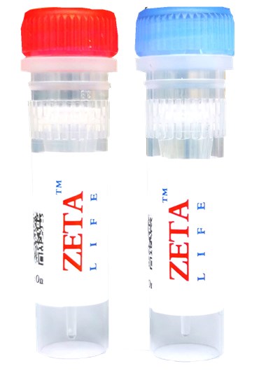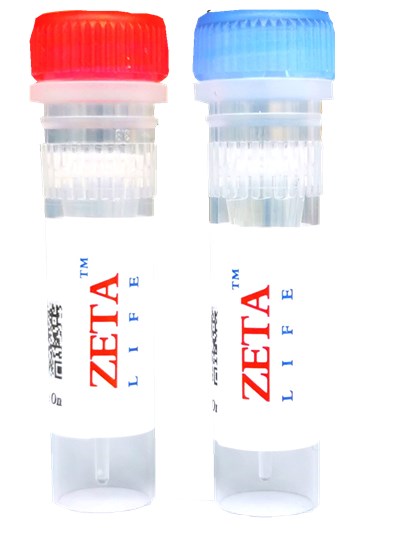

DAPI
Packing specification
Product Numbers: DA005, DA010, DA025, DA050, DA100
Specification: 5mg, 10mg, 25mg, 50mg 100mg
Storage conditions: Store at -20 ℃ away from light, valid for one year.
Shipping Condition : ambient temperature
Chemical Name: 4'''',6-Diamidino-2-phenylindole, dihydrochloride CAS: 28718-90-3
Appearance: yellow powder MW: 350.25, C16H17Cl2N5
Storage Condition : -20oC, protect from light; Shipping Condition : ambient temperature

Product Description
DAPI is an AT-sequence specific DNA intercalator that attaches to DNA at the minor groove of the double helix like Hoechst dyes. Though DAPI is not permeable through viable cell membranes, it passes through disturbed cell membranes to stain the nucleus. DAPI has a high photo-bleaching tolerance level. DAPI is utilized for the detection of mitochondrial DNA in yeast, chloroplast DNA, virus DNA, micoplasm DNA and chromosomal DNA. The excitation and emission wavelengths of DAPI-DNA complex are 360 nm and 460 nm, respectively.
Staining Procedure
1. The 1mM DAPI stock solution was prepared by adding 35mg DAPI powder into 100mL ddH2O.
2. Prepare 10-50 μM DAPI solution with PBS or an appropriate buffer.
3. Add DAPI solution with 1/10 of the volume of cell culture medium to the cell culture. Or you may replace the culture medium with 1/10 concentration of DAPI buffer solution.
4. Incubate the cell at 37oC for 10-20 min.
5. Wash cells twice with PBS or an appropriate buffer.
6. Observe the cells using a fluorescence microscope with 360 nm excitation and 460 nm emission filters.
Note: Since DAPI may be carcinogenic, extreme care is necessary during handling.
Features and Biological Applications
DAPI is a fluorescent stain that binds strongly to DNA. It is used extensively in fluorescence microscopy. Since DAPI passes through an intact cell membrane, it can be used to stain live cells besides fixed cells. For fluorescence microscopy, DAPI is excited with ultraviolet light. When bound to double-stranded DNA its absorption maximum is at 358 nm and its emission maximum is at 461 nm. One drawback of DAPI is that its emission is fairly broad. DAPI also binds to RNA although it is not as strongly fluorescent as it binds to DNA. Its emission shifts to around 400 nm when bound to RNA. DAPI''''''''s blue emission is convenient for multiplexing assays since there is very little fluorescence overlap between DAPI and green-fluorescent molecules like fluorescein and green fluorescent protein (GFP), or red-fluorescent stains like Texas Red. Besides labeling cell nuclei, DAPI is also used for the detection of mycoplasma or virus DNA in cell cultures.
For scientific research use only.


