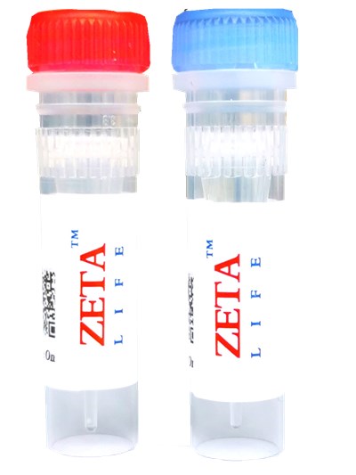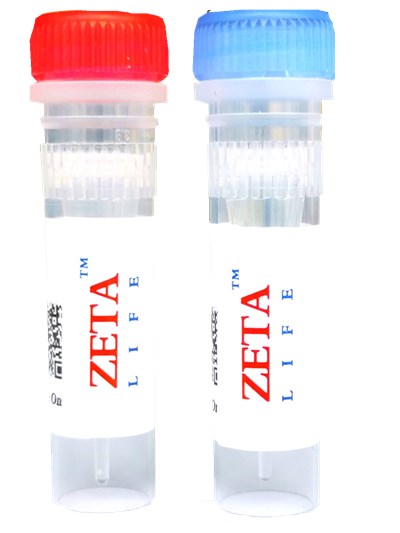

Product Numbers: DI-10, DI-25, DI-50; Specification: 10mg, 25mg, 50mg
Storage conditions: Store at 4 ° C in the dark, valid for one year. .
Appearance: red solid soluble in DMSO
λEx / λ Em (MeOH) = 491 \ 613 nm
CAS number: 114041-00-8
Molecular formula: C46 H79 IN2
Molecular weight: 787
product description
DiA is a green fluorescent dye for cell membranes. It diffuses faster in cell membranes than DiO, and is often used in conjunction with DiI for two-color labeling of cell membranes.
After DiA staining, paraformaldehyde can be fixed (methanol and other reagents are not allowed), but it is not recommended to permeabilize after staining. In addition, after fixed permeabilization (permeabilization with 0.1% TritonX-100 at room temperature), the plasma membrane can also be stained well. DiA stains fixed cells better than DiO.
Instructions
1. Preparation of DiO cell membrane staining solution
(1) Configure DMSO or EtOH storage solution: Use DMSO or EtOH for the storage solution to prepare a concentration of 1 to 5 mM.
Note: Store unused storage solution at -20 oC to avoid repeated freezing and thawing.
(2) Preparation of working solution: dilute the storage solution with a suitable buffer (such as serum-free medium, HBSS or PBS), and prepare a concentration of 1 ~
5 μM working fluid.
Note: The final concentration of the working solution is prepared according to the experience of different cells and experiments. You can find the best conditions from more than ten times the recommended concentration.
2. Staining of suspended cells
(1) Add 1 × 10⁶ / mL cell density to the working solution.
(2) The cells are cultured at 37 ℃ for 2-20 minutes, and the optimal culture time for different cells is different.
(3) Centrifuge the test tube of stained cells at 1000-1500 rpm for 5 minutes.
(4) Pour the supernatant and slowly add the pre-warmed culture medium at 37 ° C again.
(5) Repeat steps (3) and (4) more than twice.
3. Staining of adherent cells
(1) Culture the adherent cells in a sterile laboratory.
(2) Remove the coverslip from the medium, aspirate excess culture fluid, and place the coverslip in a humid environment.
(3) Add 100 μL of dye working solution to the corner of the coverslip and shake gently to make the dye evenly cover all cells.
(4) Incubate the cells at 37 ℃ for 2 to 20 minutes, and the optimal culture time for different cells is different.
(5) Absorb the dye working solution, wash the coverslip with the culture solution 2 to 3 times, cover all the cells with the pre-warmed medium each time, incubate for 5 to 10 minutes, and then absorb the medium.
4. Detection by flow cytometry
DiD, DiO, DiI, DiR and DiS stained cells can be detected by the classic FL1, FL2, FL3 and FL4 flow cytometers, respectively.
For scientific research use only.


